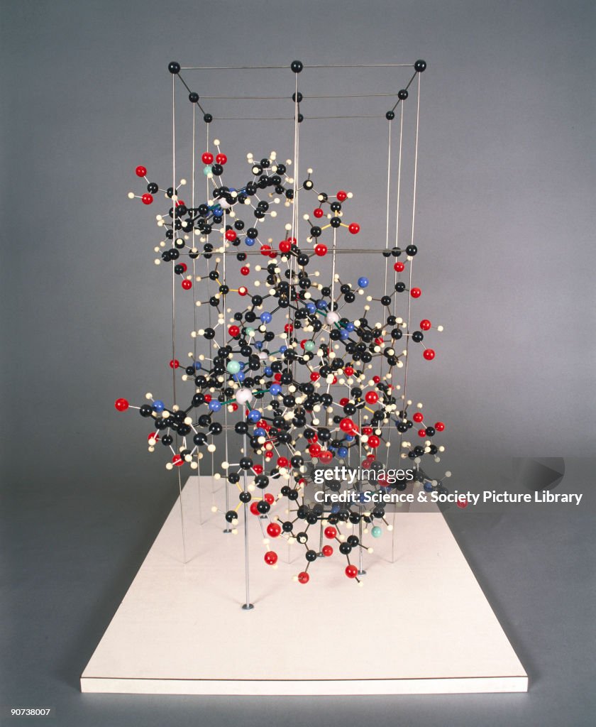
Vitamin B12 crystal structure model, 1957-1959.
UNITED KINGDOM - JULY 02: This crystal structure model, made for the X-ray crystallographer Dorothy Crowfoot Hodgkin, shows the structure of the hexacarbocylic acid fragment of Vitamin B12. It was displayed at the Brussels Universal Exhibition (�Expo�) in 1958. The full structure of this complex and large vitamin was determined by Hodgkin using X-ray crystallography in 1957, before chemists were able to work out using chemical means, and this model is a result of her work. Vitamin B12 is produced by micro-organisms in the gut. The richest natural source is raw liver. Deficiency leads to pernicious anaemia, which is the poor development of red blood cells resulting in the possible degeneration of the spinal chord. Sufferers develop extensive bruising and recover slowly from even minor injuries. (Photo by SSPL/Getty Images)

PURCHASE A LICENSE
How can I use this image?
€300.00
EUR
Getty ImagesVitamin B12 crystal structure model, 1957-1959., News Photo Vitamin B12 crystal structure model, 1957-1959. Get premium, high resolution news photos at Getty ImagesProduct #:90738007
 Vitamin B12 crystal structure model, 1957-1959. Get premium, high resolution news photos at Getty ImagesProduct #:90738007
€475€115
Getty Images
In stockPlease note: images depicting historical events may contain themes, or have descriptions, that do not reflect current understanding. They are provided in a historical context. Learn more.
DETAILS
Restrictions:
Contact your local office for all commercial or promotional uses.
Credit:
Editorial #:
90738007
Collection:
SSPL
Date created:
July 02, 1998
Upload date:
License type:
Release info:
Not released. More information
Source:
SSPL
Object name:
10311590
Max file size:
2875 x 3504 px (9.58 x 11.68 in) - 300 dpi - 2 MB