Trachea mucous membrane - stock illustration
Trachea mucous membrane. Coloured scanning electron micrograph (SEM) of a fractured mucous membrane of the trachea (wind pipe), showing the epithelium and underlying connective tissue. The upper epithelial surface is covered in a mixture of secretory cells (brown) and ciliated cells (pink). The tall columnar epithelium (green) is exposed with some nuclei visible, and the underlying connective tissue (lamina propria) is seen at bottom. Magnification: x1000 when printed at 10 centimetres wide.

Get this image in a variety of framing options at Photos.com.
PURCHASE A LICENSE
All Royalty-Free licenses include global use rights, comprehensive protection, simple pricing with volume discounts available
€300.00
EUR
Getty ImagesTrachea Mucous Membrane High-Res Vector Graphic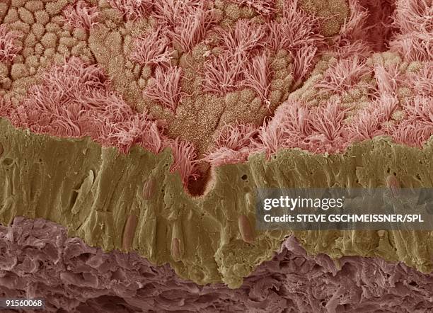 Download premium, authentic Trachea mucous membrane stock illustrations from Getty Images. Explore similar high-resolution stock illustrations in our expansive visual catalogue.Product #:91560068
Download premium, authentic Trachea mucous membrane stock illustrations from Getty Images. Explore similar high-resolution stock illustrations in our expansive visual catalogue.Product #:91560068€300€40
Getty Images
In stockDETAILS
Credit:
Creative #:
91560068
License type:
Collection:
Science Photo Library
Max file size:
4955 x 3579 px (16.52 x 11.93 in) - 300 dpi - 4 MB
Upload date:
Release info:
No release required
Categories:
- SEM,
- Mucosal Membrane,
- Wineglass,
- Human Gland,
- Illustration,
- Mucus,
- Biological Cell,
- Bone Fracture,
- Epithelium,
- Scanning Electron Microscope,
- Anatomy,
- Biology,
- Biomedical Illustration,
- Cilium,
- Color Image,
- Connective Tissue,
- Cross Section,
- Healthy Lifestyle,
- Horizontal,
- Human Body Part,
- Human Tissue,
- Human Trachea,
- Illustration Technique,
- Lamina Propria,
- Microbiology,
- Microvillus,
- No People,
- Nucleus,
- Respiratory Tract,
- Secretory Cell,
- Simplicity,